Open the same H&E slide in two viewers. One pink is a little bluer, the other a little warmer. Which one is right?
The scanner knows. Its camera sees colour its own way, and many scanners store a small description of that camera inside the slide: an ICC colour profile [1]. It says how to turn the numbers in the file into the colour you should see.
The Find Nuclei Viewer now reads that profile and draws the slide with it. In your browser, with nothing to install.
One button
Open a slide. If its file carries a profile, the viewer uses it right away. The ICC button sits next to RGB, BW and INV, and turns it off and on, so you can compare.
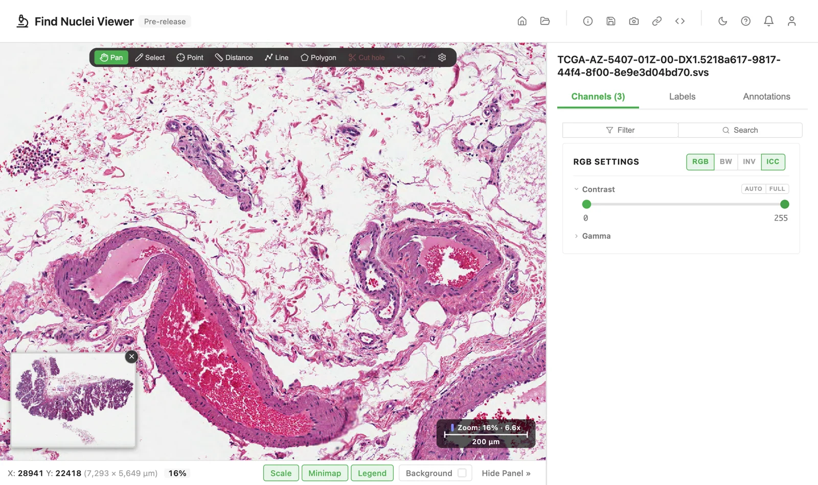
No profile in the file? The button is greyed, and the slide looks exactly as before.
Contrast and gamma still work as always. The profile comes first, your adjustments go on top. Turn ICC off, and a shared link keeps it off for the person who opens it.
Before and after
Aperio
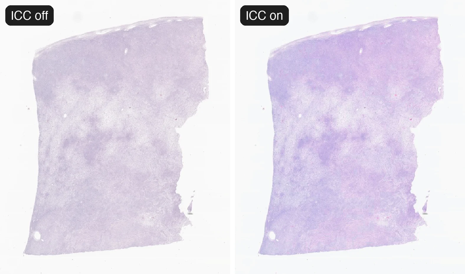
Every Aperio SVS we tried that came straight from the scanner carries a profile. Only exported copies lacked one. Here is a colon adenocarcinoma slide from TCGA [2], straight from the Genomic Data Commons. Left without the profile, right with it.
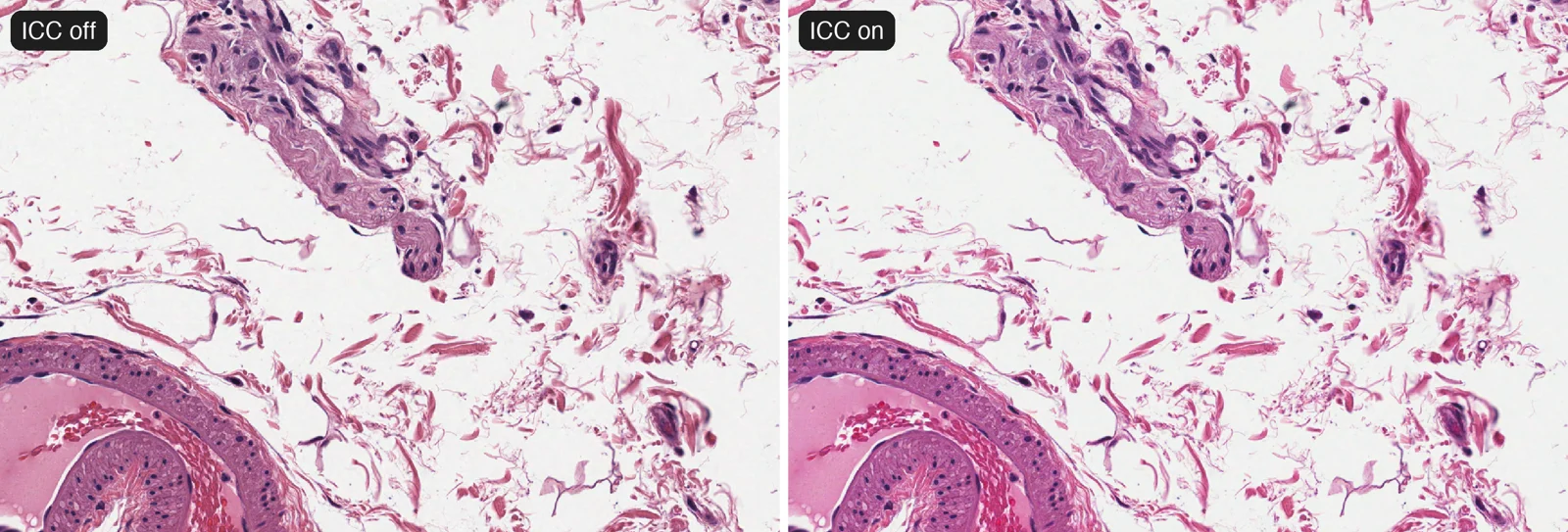
The eosin turns warmer and pinker. The violet cast leaves the tissue. Open this spot yourself and click ICC.
Hamamatsu
A NanoZoomer S20MD slide in DICOM [3][4] carries a profile that says its pixels are linear light. A colour-managed viewer then brightens the midtones a lot. The result is much paler than the raw pixels.
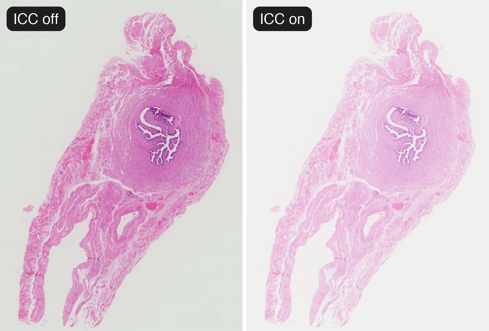
That is what the file says, and the viewer follows it. If you prefer the raw pixels, one click on ICC shows them.
3DHISTECH
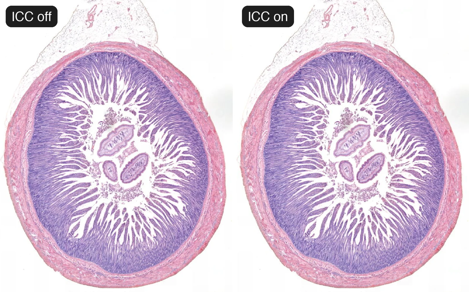
A Pannoramic 1000 slide in DICOM [3] carries a modern version 4 profile built on sRGB. The change is small: shadows sit a little deeper.
Leica
An Aperio GT450 slide in DICOM [3] carries a profile of 13 MB. Here it deepens the violet of the hematoxylin.
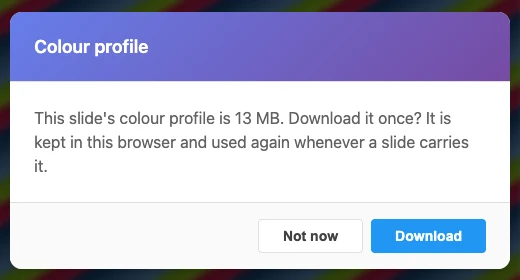
From your disk, the viewer simply reads the profile. By address, 13 MB is a lot to fetch before the first picture, so the viewer asks first. Confirm once, and the profile is kept in your browser. Every later slide with the same profile draws with it at once.
True to the profile
A profile is a recipe, and a recipe can be followed badly. We checked the viewer against Little CMS, a widely used colour engine [5], on the real profiles above. The difference is below what the eye can see.
It is quick too. The profile is read together with the slide’s layout, in the same requests. It is kept in your browser, so the next open fetches no profile at all, and a published slide index carries it for everyone.
Limitations
- OME-TIFF and Akoya QPTIFF files do not use a profile yet.
- A Leica SCN slide with several regions uses the profile of the first one.
- The embedded viewer for publications draws without profiles for now.
- The profile changes the picture on your screen, never the file. Analysis reads the numbers the scanner wrote.
Try it now
Open the TCGA colon slide and click ICC a few times. Or open your own slide: drop the file on the viewer, or paste its address. The Channels guide explains the button. For more on slides in the browser, read Whole Slides in the Browser.
References
[1] Specification ICC.1:2022, Image technology colour management. International Color Consortium (2022). www.color.org/icc_specs2
[2] Grossman RL, Heath AP, Ferretti V, et al. Toward a Shared Vision for Cancer Genomic Data. New England Journal of Medicine 375, 1109-1112 (2016). doi.org/10.1056/NEJMp1607591. The results shown here are in whole or part based upon data generated by the TCGA Research Network: www.cancer.gov/tcga. Slide TCGA-AZ-5407-01Z-00-DX1.
[3] OpenSlide test data. Carnegie Mellon University. openslide.cs.cmu.edu/download/openslide-testdata. Aperio CMU-1, DICOM Hamamatsu-2, 3DHISTECH-1 and Leica-4, CC0.
[4] DICOM PS3.3, section C.11.15, ICC Profile Module. NEMA. dicom.nema.org/medical/dicom/current/output/chtml/part03/sect_C.11.15.html
[5] Little CMS. Marti Maria. www.littlecms.com