Someone sent you a study. Forty .svs files, and you just want to look at them.
So you download a desktop viewer. You install it, or you file an IT ticket because the laptop is locked down. You make a project. You download the slides, a gigabyte or two each, and wait for tens of gigabytes. You add them to the project. Then you want a colleague’s opinion, and they start the same list from the top.
Desktop tools are great for analysis. For looking at slides, it is a lot of homework.
Here is the short version: open them in a web page.
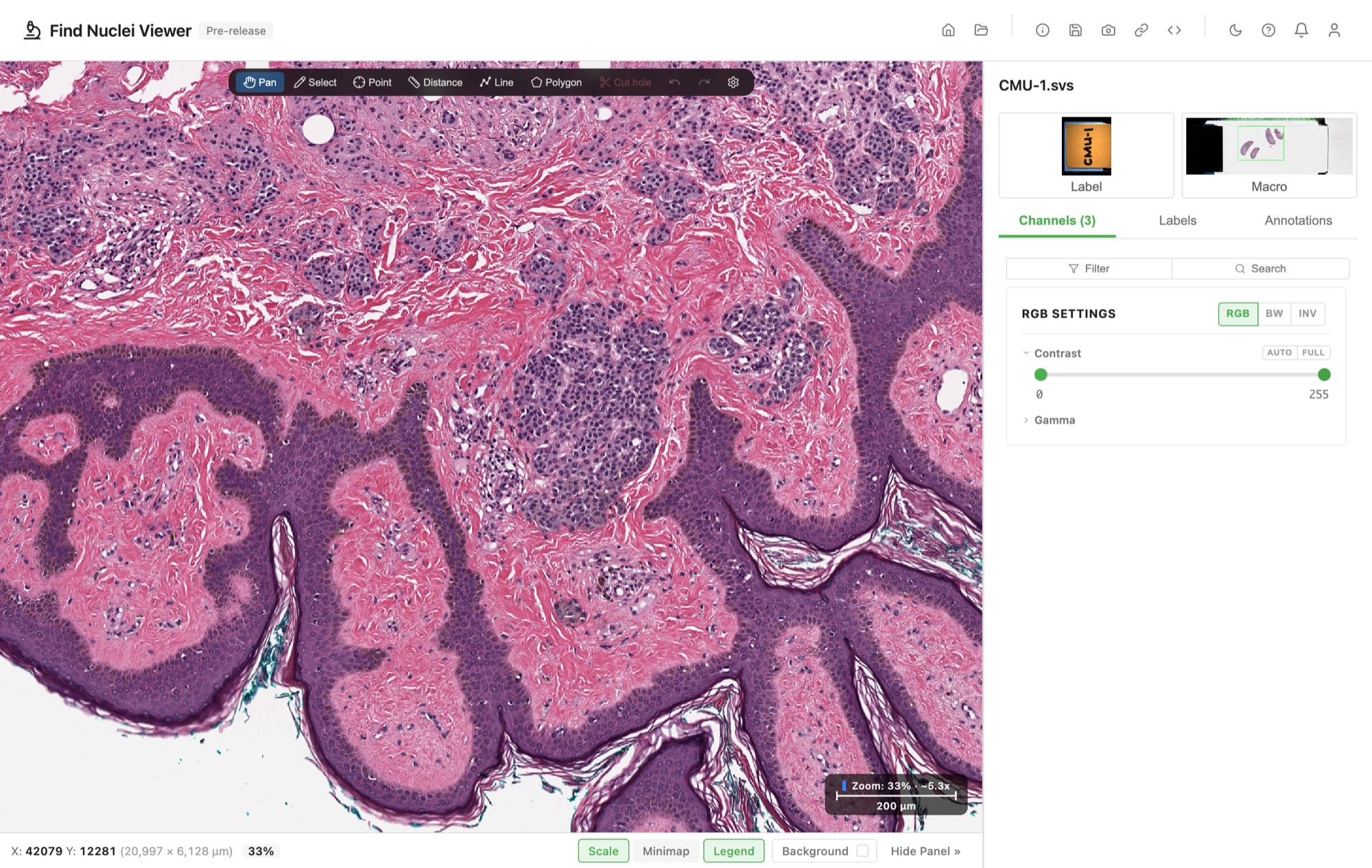
Three steps, no homework
- Go to find-nuclei.github.io.
- Drop your slide on the page.
- Look.
A whole study? Drop the folder instead, and pick a slide from the list. Folders open in Chrome and Edge.
No slide at hand? Open a lymph node slide with its metastases outlined, from the CAMELYON16 challenge on AWS Open Data [1]. One click, nothing downloaded.
Nothing to install, nothing to set up, no project. The slide stays on your computer, and no server ever sees a pixel. Zoom in until you see single cells. The viewer reads only the part on screen, so it stays fast all the way down.
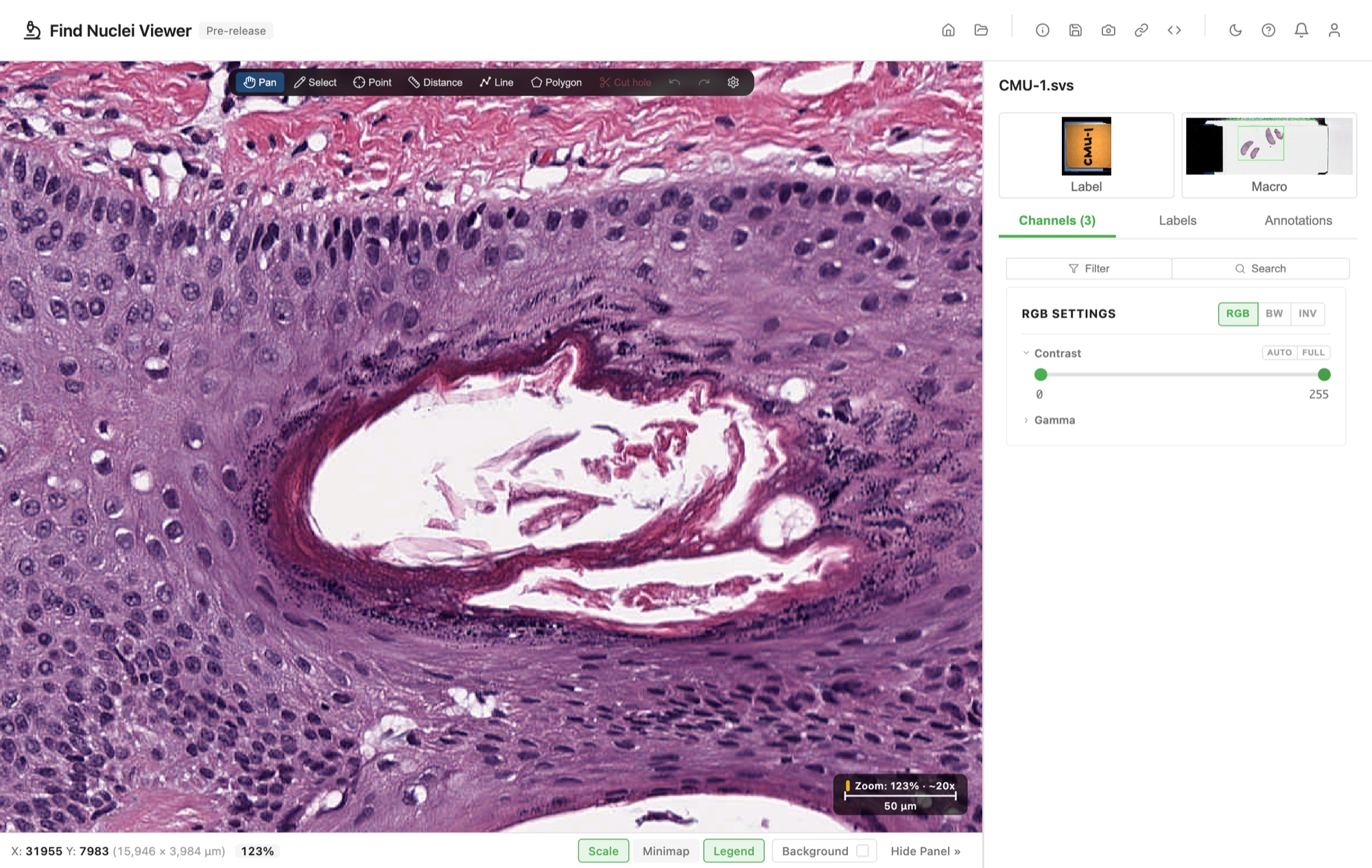
The slide in these pictures is CMU-1, a free test slide from OpenSlide [2].
No download either
Are the slides in a bucket or on a server? Then you do not download them at all. Paste an address, https://, s3://, or gs://, and that slide opens. Your browser fetches the tiles you look at, and nothing else. Forty slides cost you what you actually look at, not 60 GB of waiting.
Private storage works too: a private link or a token, and the slide opens straight from your storage. Private Images and Slides shows how.
Side by side
| Desktop software | Find Nuclei Viewer | |
|---|---|---|
| Install | A desktop app | Nothing, a browser tab |
| Get the slides | Download them all first | Drop one, or paste its address |
| Set up | A project, then add the images | None |
| Show a colleague | They install it and get the file too | Send a link |
| Annotations | GeoJSON | The same GeoJSON, both ways |
Keep your desktop work
You do not have to choose. Annotations travel both ways in GeoJSON, with QuPath and other tools. Classes keep their names and colours, and shapes land in the same place.
Draw in QuPath, export, drop the file here. Or draw here, export, drag it onto the slide in QuPath. Annotations from Aperio ImageScope and NDP.view open too, and the same GeoJSON goes on to Visiopharm [3] and 10X Genomics Xenium Explorer [4].
One link for a second opinion
Here is the part that ends the hard drive in the post.
Put the slide and its annotation file next to each other in your storage. Paste both into the viewer and copy the link. Your colleague clicks it. The slide opens in their browser with your annotations on it.
They need a browser. Not a licence, not an installer, not an IT ticket.
This is what they see: a CAMELYON16 slide in a public bucket, with the pathologists’ tumour outlines from the file beside it.
One viewer for SVS, NDPI, SCN, MRXS, QPTIFF, and DICOM
| Scanner | File | How it opens |
|---|---|---|
| Aperio | .svs | One file |
| Hamamatsu | .ndpi | One file, over 4 GB too |
| Leica | .scn | One file, every scanned region |
| Philips | .tiff | One file |
| Akoya | .qptiff | One file, multiplex fluorescence included |
| 3DHISTECH | .mrxs | The file and its folder, or a zip |
| DICOM WSI | .dcm | A folder, or a zip |
Each one opens from your disk, or by its address. Supported Formats has a guide for each, and the microscopy formats too.
| Annotations | Import | Export |
|---|---|---|
| GeoJSON (QuPath, Visiopharm [3], Xenium Explorer [4]) | Yes | Yes |
| OME-XML | Yes | Yes |
| Aperio ImageScope XML | Yes | |
Hamamatsu NDP.view .ndpa | Yes | |
| ASAP XML (CAMELYON16) | Yes |
Exchanging Annotations shows each tool step by step.
Limitations
- Analysis stays on your desktop. This is for looking, measuring, annotating, and sharing. For cell detection and classifiers, keep your analysis tools.
- SVS over 4 GB. Large Aperio files use BigTIFF, and the viewer does not read those yet. It tells you so, by name.
- Folders need Chrome or Edge. MRXS and DICOM slides are folders. In Firefox and Safari, zip them without compression first.
Try it now
Close the IT ticket. Open the slide.
- A lymph node with metastases, outlined by the CAMELYON16 pathologists [1]
- A colon adenocarcinoma from The Cancer Genome Atlas, straight from the NCI Genomic Data Commons [5]
Slide refuses to open? Tell us at info@find-nuclei.com.
References
[1] Litjens G, Bandi P, Ehteshami Bejnordi B, et al. 1399 H&E-stained sentinel lymph node sections of breast cancer patients: the CAMELYON dataset. GigaScience 7(6), giy065 (2018). doi.org/10.1093/gigascience/giy065. CC0.
[2] OpenSlide test data, Aperio. Carnegie Mellon University. openslide.cs.cmu.edu/download/openslide-testdata/Aperio
[3] Working with Layer Data. Visiopharm Manual, version 2025.08.1. Visiopharm. cpwebapps.azurewebsites.net/docs/2025.08.1/database/layer_data
[4] Selecting multiple regions of interest. Xenium Explorer documentation. 10X Genomics. www.10xgenomics.com/support/software/xenium-explorer/latest/tutorials/xe-selecting-multiple-regions-of-interest
[5] Grossman RL, Heath AP, Ferretti V, et al. Toward a Shared Vision for Cancer Genomic Data. New England Journal of Medicine 375, 1109-1112 (2016). doi.org/10.1056/NEJMp1607591. Data generated by the TCGA Research Network.