Your CellVoyager finished the plate. It left a measurement folder behind: a MeasurementDetail.mrf, a .mes, a MeasurementData.mlf, and tens of thousands of TIFFs. Now you want to see well C/7.
Usually that means the acquisition PC, or a conversion job before you see a single cell.
Or you paste the folder address into your browser.
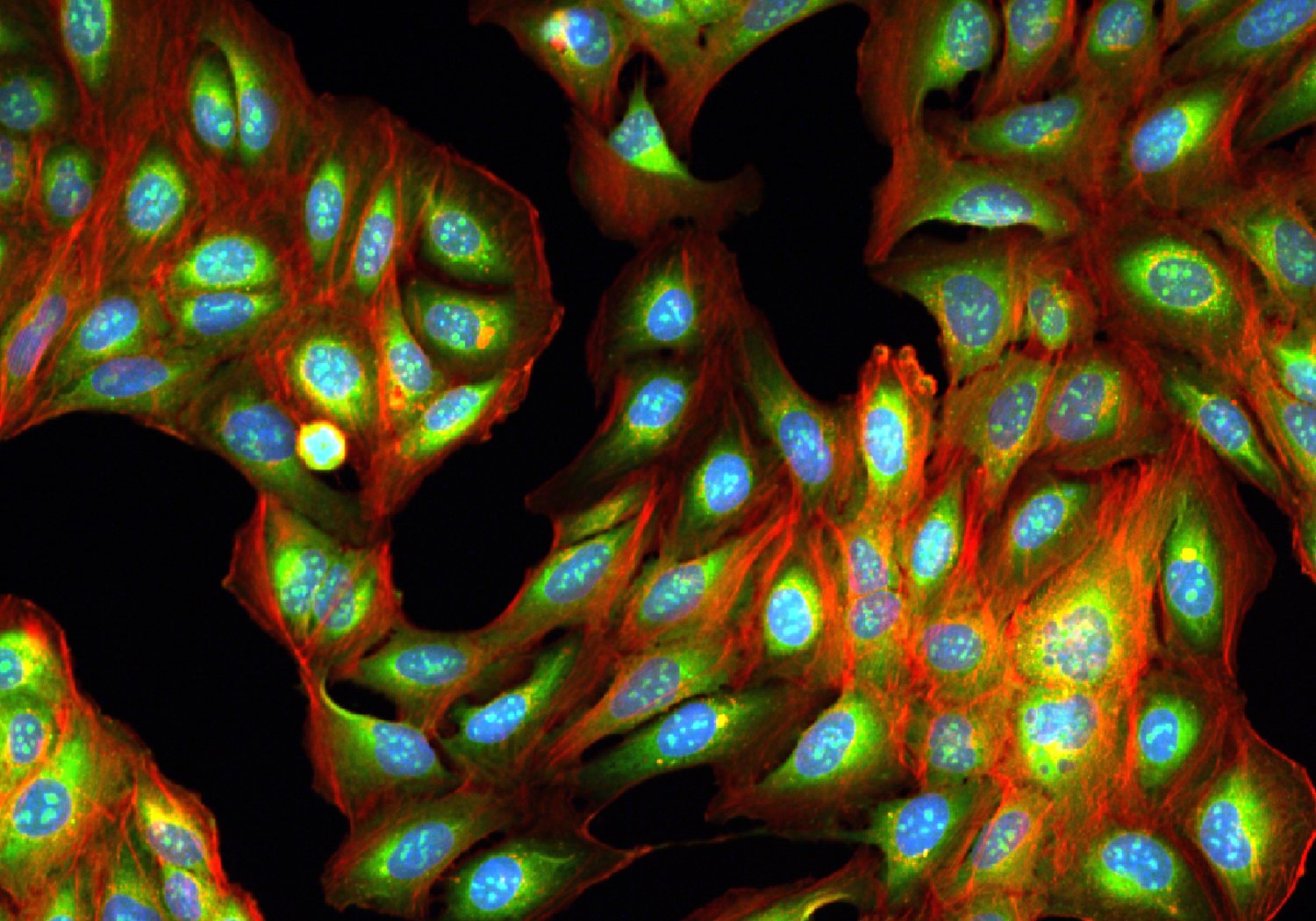
Last week the Find Nuclei Viewer learned to open Opera Phenix plates. This week it opens plates from the Yokogawa CellVoyager CV8000 and CV7000, straight from the measurement folder. The TIFFs stay where they are. Free to use, nothing to install, nothing uploaded.
Try it on a real plate
The Cell Painting Gallery [1] holds the JUMP Cell Painting dataset [2]: Cell Painting images of U2OS cells under about 136,000 chemical and genetic perturbations, imaged by labs across the JUMP consortium. More than 1,000 of its plates come from a CellVoyager.
Here is one of them, plate 1053601756, a 384-well CV8000 plate:
Open plate 1053601756 in the viewer
That link is the plate’s S3 address, plus three stains switched on: DNA in blue, ER in green, actin and Golgi in red. The address alone works too:
s3://cellpainting-gallery/cpg0016-jump/source_2/images/20210607_Batch_2/images/1053601756/The plate opens as soon as its first well is in. The rest of the image list finishes in the background while you look around.
What you get
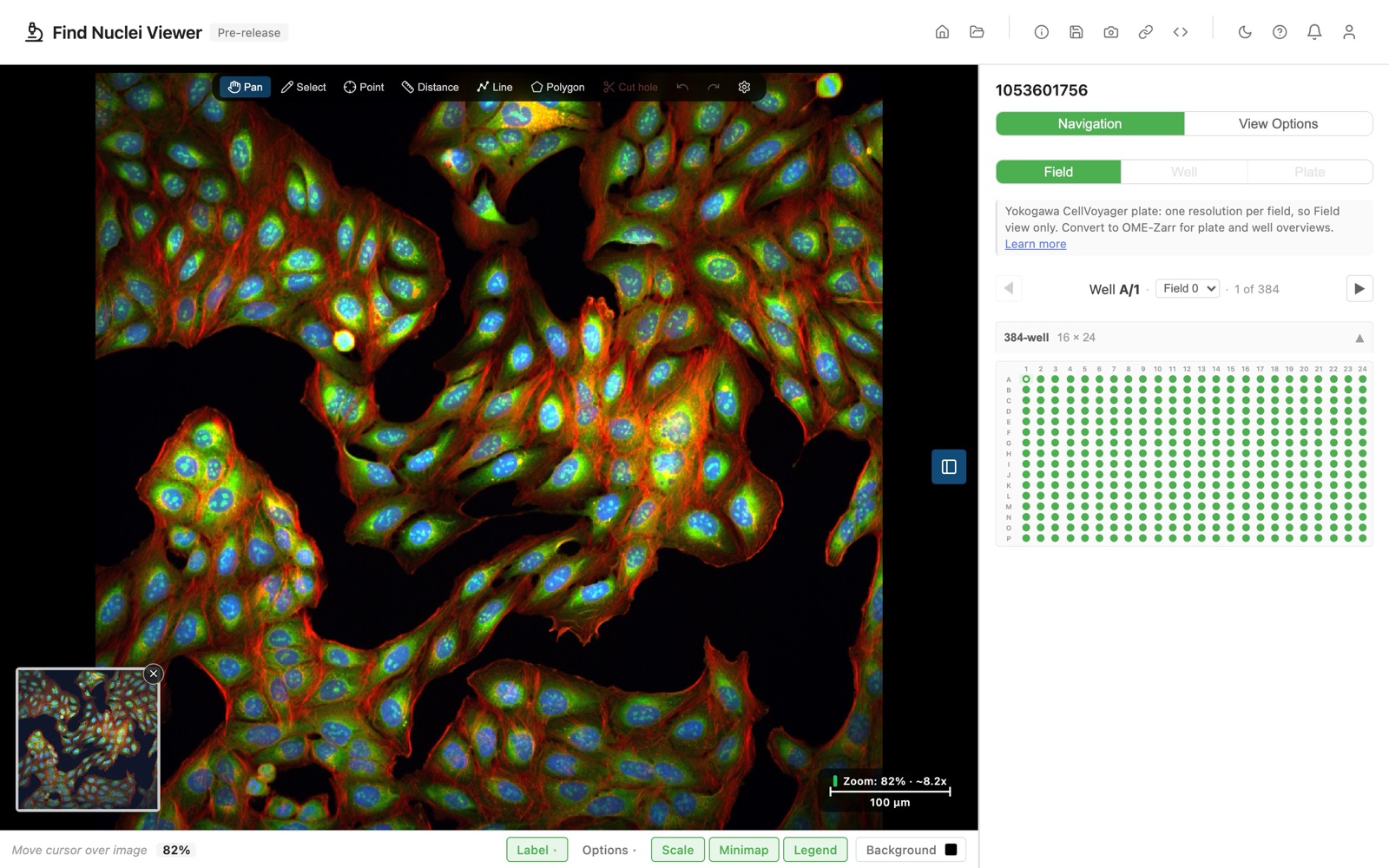
- The whole plate. Every imaged well and every field. Step through them with the well navigator, or jump to any well in the picker.
- Your channel names. They come from the
.mesfile: the dye, the filter and the light source of each channel. - Real units. The scale bar reads the pixel size from the measurement.
- Share links. Copy the link, and a colleague opens the same well, field, and channels in their browser.
- A faster second visit. The viewer remembers what it learned about the plate, so the next open takes well under a second.
Three ways to open a plate
- From a URL. Paste the measurement folder, or the address of its
MeasurementDetail.mrforMeasurementData.mlf.https://ands3://both work. - From your disk. Open the measurement folder in Chrome or Edge. The files never leave your computer.
- From a plate manifest. “Save plate manifest” writes a small JSON file that maps every image on the plate. Put it next to the images, and the plate opens without reading the metadata files again.
The Yokogawa CellVoyager plates guide walks through each one.
Harmony V6 too
Harmony V6 writes its index as Index.xml, not Index.idx.xml. That one opens now as well, the same way as a V5 export. The Opera Phenix plates guide covers both versions.
How much of JUMP opens now
By our count of the JUMP plates in the Cell Painting Gallery, Opera Phenix and CellVoyager plates together make up about four in five. All of those now open in the browser, image by image, with nothing downloaded first.
Limitations
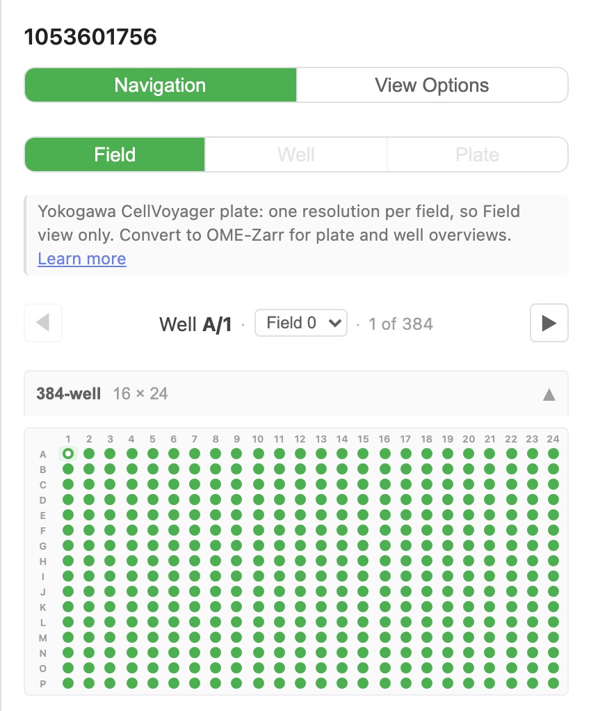
A CellVoyager writes each field as plain TIFFs at one resolution. That sets the trade-offs:
- Field view, one field at a time. A plate or well overview would mean loading every image on the plate. The Plate and Well buttons say why.
- Brightfield shows plane 1. The CV8000 records several Z planes for brightfield. The viewer shows the first.
- Images show as recorded. No flat-field correction yet.
- Some folders do not open yet. The Yokogawa CQ1 writes a different set of files, and folders that hold TIFFs without the metadata files have nothing to read.
Need plate overviews, or fast browsing on a slow link? Convert the plate to OME-Zarr. For checking a plate, the TIFFs as they are work fine.
What comes next
Looking at the plate is step one. Step two is analysis: segment the cells, measure them, and see the results on top of the same images, as in our HCS analytics post.
Have CellVoyager plates of your own? Paste one in. If one does not open, send us a note at info@find-nuclei.com. We want to see it.
Which HCS format should we add next?
Opera Phenix and CellVoyager open today. Supported Formats lists everything the viewer reads. Which instrument should be next? Tell us at info@find-nuclei.com.
TL;DR
- Find Nuclei Viewer opens Yokogawa CellVoyager CV8000 and CV7000 plates from the measurement folder:
MeasurementDetail.mrf,.mes,MeasurementData.mlf, plus the TIFFs. - Harmony V6 (
Index.xml) opens too, next to V5. - Open from a URL (
https://ors3://), from your disk in Chrome or Edge, or from a saved plate manifest. - No conversion, no upload. Free to use.
- One field at a time: no plate overview. Convert to OME-Zarr when you need overviews.
- Try it on JUMP plate 1053601756 from the Cell Painting Gallery.
References
[1] Weisbart E, Kumar A, Arevalo J, Carpenter AE, Cimini BA, Singh S. Cell Painting Gallery: an open resource for image-based profiling. Nature Methods 21, 1775-1777 (2024). doi.org/10.1038/s41592-024-02399-z
[2] Chandrasekaran SN, Ackerman J, Alix E, et al. JUMP Cell Painting dataset: morphological impact of 136,000 chemical and genetic perturbations. bioRxiv (2023). doi.org/10.1101/2023.03.23.534023