You segmented 163 cells. In 2D the outlines look fine, plane by plane.
But did the segmentation follow each cell through the stack? Are two nuclei stuck together in z? Does that boundary sit on the membrane, or a few planes off?
Those are 3D questions. The Find Nuclei Viewer now shows your label images in 3D, in the same browser tab. Free to use, nothing to install, nothing uploaded.
Try it on a real stack
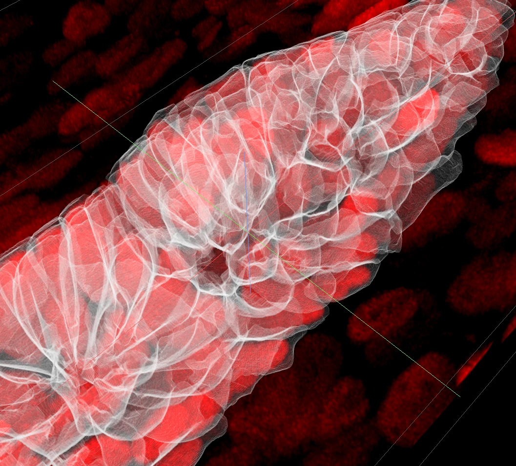
This is the zebrafish lateral line primordium from the Image Data Resource, study idr0079 [1], with its cell segmentation. Every cell wall is a thin glass outline over the nuclei. You see the label and the image at once.
Drag to turn it. Scroll to zoom. The settings: label as Volumetric, Contour only, white, width 1, opacity 0.2. Nuclei channel only, MIP.
Check your segmentation against the membranes
Put the membrane channel on and draw the label as a thin red outline. Where a red wall lies on a green membrane, the segmentation is right. Where they drift apart, you found a problem, and you can see in which plane.
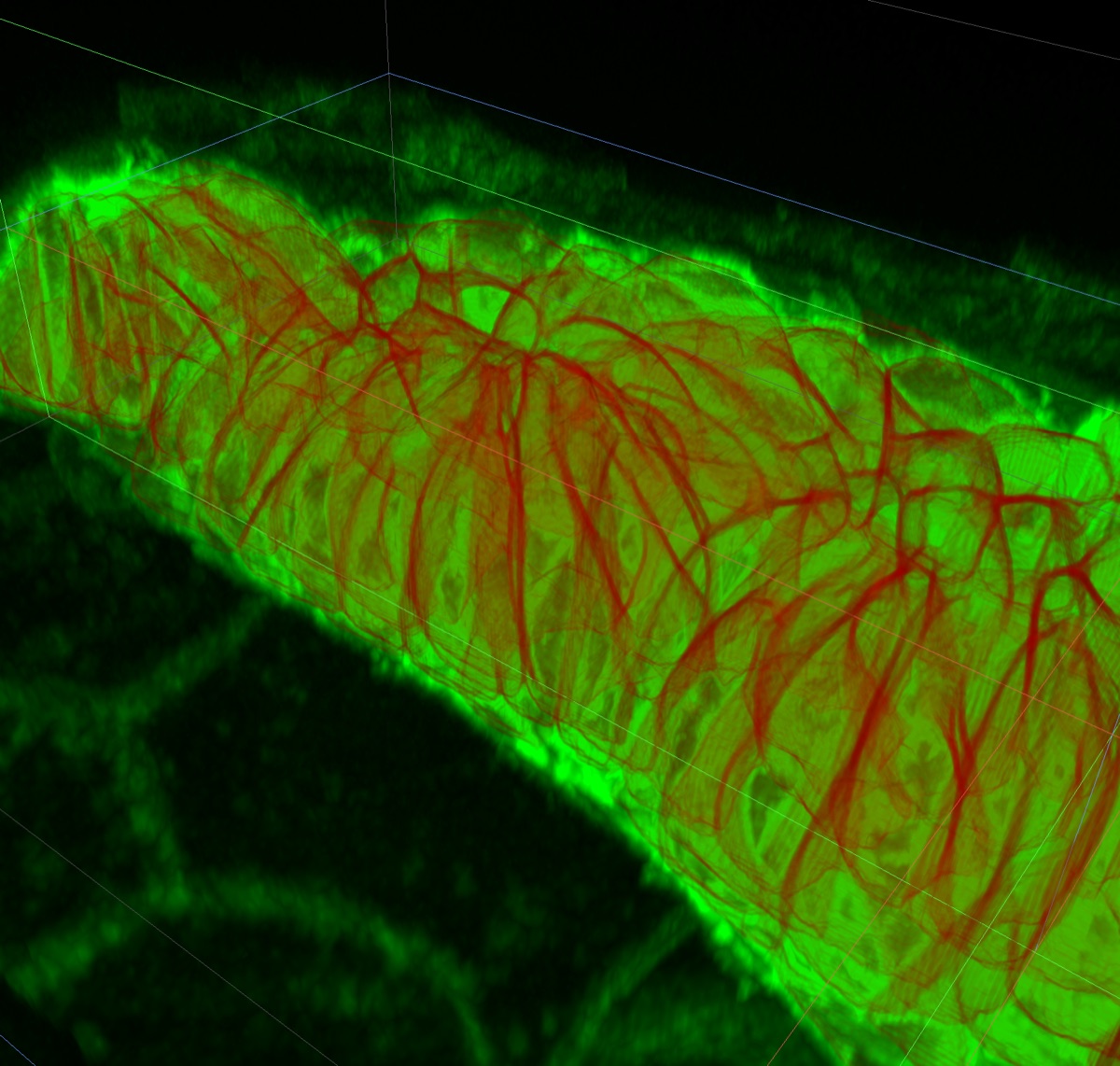
The settings: membranes in green, MIP. Label Contour only, red, width 1, opacity 0.2.
See inside each object
Labels do not have to cover the image. Draw each object as a translucent shell, and the channels inside stay visible.
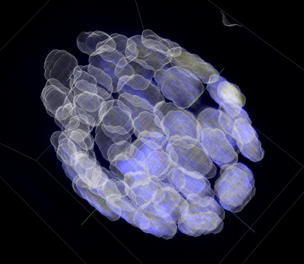
Mouse nuclei from study idr0062 [2]. Each nucleus is a white glass shell around its lamina, in blue.
The settings: label Contour only, white, opacity 0.35. Lamin B1 in blue, contrast 0 to 2801. MIP, High Quality on.
Two looks: Surface and Volumetric
- Surface draws each object as a closed, smooth surface. It is the default. Good for counting, shapes and objects that touch.
- Volumetric colours the volume itself, blended with the channels. Good for outlines over the image, as in the views above, and for labels with very many objects.
Both use the colours you set in 2D: one solid colour, random colours, or a heatmap of a measurement.
Three settings do most of the work:
- Fill or Contour. Contour shows the image inside each object.
- Width, from 1 to 5. A thicker outline reads better from far away.
- Opacity. Low opacity turns a label into glass over the channels.
Bring your own labels
- Open your OME-Zarr image, as you do today.
- Your labels come with it: the ones embedded in the image, or add more with Add Local or Add URL in the Labels tab.
- Click 3D.
Then copy the link. It carries the labels, including the ones you added with Add URL, with their colours and look. Your colleague opens the same view.
The 3D Rendering guide covers every setting.
Limitations
Straight talk on limitations:
- A desktop browser with WebGPU. Recent Chrome and Edge have it.
- Surfaces take a moment. The viewer builds them while you look, and counts them in the corner of the 3D view. The 163 cells above take about ten seconds. Labels with many thousands of objects take longer; Volumetric shows them at once.
- Four Volumetric labels at once. Surface labels do not count toward the four.
- Thick z-planes show as steps. When the z-step is much larger than the pixel size, label walls can show fine stripes, one per plane. That is the data, drawn honestly. Smoothing that takes the voxel shape into account is on our list.
Have a segmentation that looks wrong in 3D, or does not open? Let us know at info@find-nuclei.com.
TL;DR
- Label images now show in 3D, next to the image.
- Every object as a smooth surface, or as an outline over the channels.
- Contour, width and opacity turn labels into glass, so you see the label and the image at once.
- Copy a link, and your labels come with it.
- No install, no upload. Free to use.
References
[1] Image Data Resource, study idr0079, zebrafish lateral line primordium. idr.openmicroscopy.org/study/idr0079. Paper: doi.org/10.7554/eLife.55913. CC BY 4.0.
[2] Image Data Resource, study idr0062, mouse nuclei. idr.openmicroscopy.org/study/idr0062. Paper: Blin et al. 2019, doi.org/10.1371/journal.pbio.3000388. CC BY 4.0.