You want to know what happens in the middle of the tissue. The outer cells are in the way.
Or your slide is 51,000 pixels wide, and 3D shows it as a blur, because no graphics card holds that much in one piece.
Same fix for both: draw a box. The Find Nuclei Viewer shows what is inside it alone in 3D, at the finest detail that fits, with your labels cut along with it. Free to use, nothing to install, nothing uploaded.
Open a tissue like a book
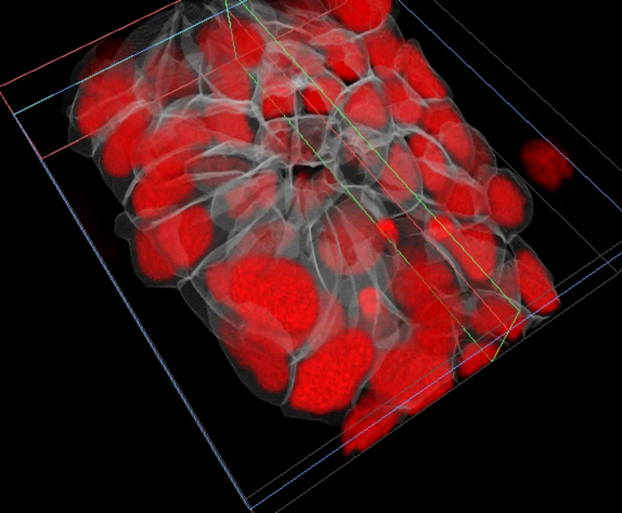
This is a rosette in the zebrafish lateral line primordium, from study idr0079 [1]. The box takes 54 of the 141 z-sections and about 11 MB, at full detail. The red is the nuclei. The white lines are the cell walls, drawn from the segmentation and cut with the image. They converge on the centre of the rosette.
A thinner slab of the same tissue, seen from the side: each nucleus sits in its cell, cut clean by the edge of the box.
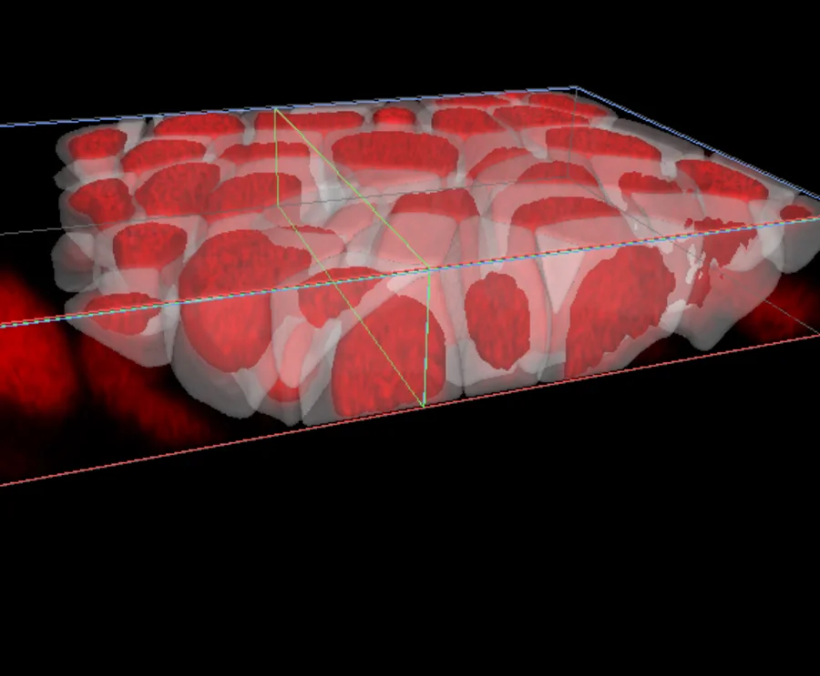
A slab this thin is a small read, and the box tells you how small before it loads.
See through the top layers
Composite shows near things in front of far things, so the surface hides the inside. A thin slab takes the surface away.
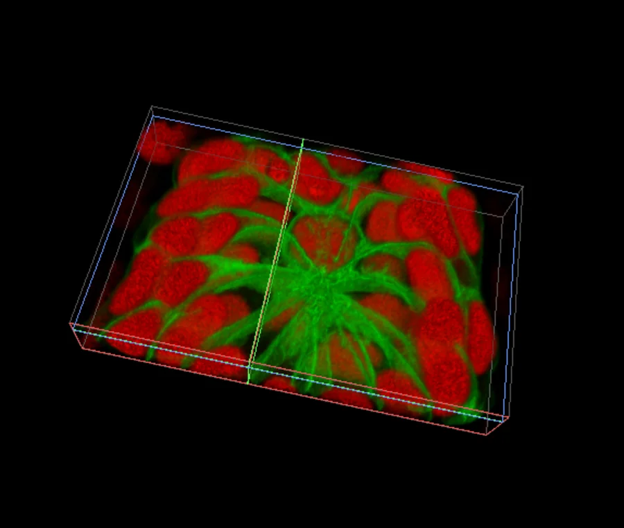
Open a thin slab of membranes and nuclei
The membranes run into the centre of the rosette like spokes. In the whole volume, the cells on top cover them.
Know what you get before you read it
Choose Region on the toolbar and drag a box on the XY, XZ or YZ panel. Nothing loads while you draw. The box tells you what 3D will give you:
Full detail · 0.21 µm · about 130 MB
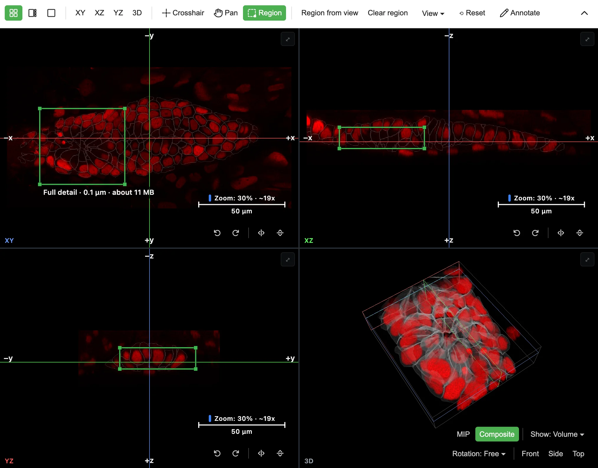
Green is full detail. Amber means only coarser detail fits, and it says which. Red means nothing fits at this size. Make the box smaller until it turns green, and you have the largest area at full detail.
Let go. The 3D panel empties, then fills with that box.
The numbers count the channels you show, your labels, your 3D memory setting and your graphics card. What the box promises is what you get.
The same box for very large slides
A Xenium slide of liver tissue [2] is 51,000 by 51,000 pixels. As a whole it shows in 3D at 1/32 detail. A box of 2,048 by 2,048 pixels shows at full detail, single nuclei and all, for about 130 MB of a slide that holds about 78 GB before compression.
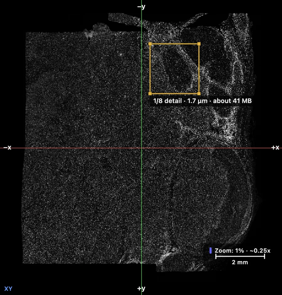
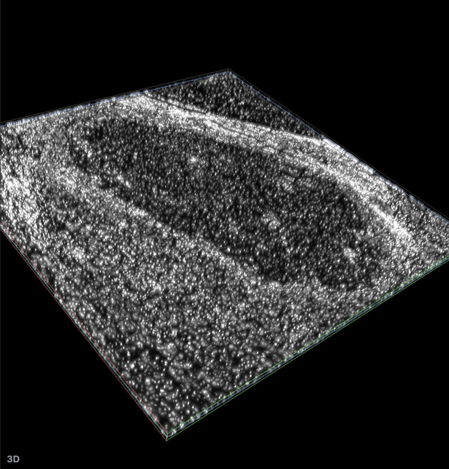
Left, the whole slide with a box on its top right part. Right, that box in 3D, at 1/8 detail for about 41 MB. The same shapes show in both, so you can see where you are. DAPI, BioImage Archive S-BIAD2339, CC0 [2].
Open a box of 2,048 by 2,048 pixels at full detail, or the larger box above
It works with no pyramid too. A Zeiss CZI file with one resolution opens in a region at full detail, because a box reads only the part of the image it holds.
Faster: use what you are looking at
Zoom in on a plane and click Region from view. The box takes what the XY panel shows, and the z-sections from XZ or YZ if you zoomed that panel last.

Clear region goes back to the whole image.
Then copy the link. It carries the box, the camera and your labels, so a colleague opens the same cut.
The 3D Region guide covers every setting.
Limitations
Straight talk on limitations:
- A desktop browser with WebGPU. Recent Chrome and Edge have it.
- Boxes are straight. They follow the image axes. No oblique cuts yet.
- One box at a time. Only the box shows in 3D. The planes keep the whole image around it.
- A box larger than the graphics card allows shows at coarser detail, or offers to read it at less detail in X and Y. It tells you which.
- A label saved at another size than the image can sit up to one of its larger voxels off.
Got an image a box cannot open? Let us know at info@find-nuclei.com.
TL;DR
- Draw a box on any plane, and 3D shows that part alone, at the finest detail that fits.
- The box tells you the detail and the size of the read before anything loads.
- Labels and surfaces are cut along with the image.
- Copy a link, and a colleague opens the same cut.
- No install, no upload. Free to use.
References
[1] Image Data Resource, study idr0079, zebrafish lateral line primordium. idr.openmicroscopy.org/study/idr0079. Paper: doi.org/10.7554/eLife.55913. CC BY 4.0.
[2] BioImage Archive, S-BIAD2339, spatial in situ transcriptomics of post-transplant HCC recurrence using 10x Genomics Xenium. doi.org/10.6019/S-BIAD2339. CC0.