You imaged a z-stack. 142 planes. The 2D view shows them one at a time, and you scroll up and down and try to keep the shape in your head.
Is that one nucleus or two stacked on top of each other? Does the membrane signal hold all the way down? Did your segmentation follow the cell in z, or give up halfway?
Those are 3D questions. Now you can answer them in the same browser tab.
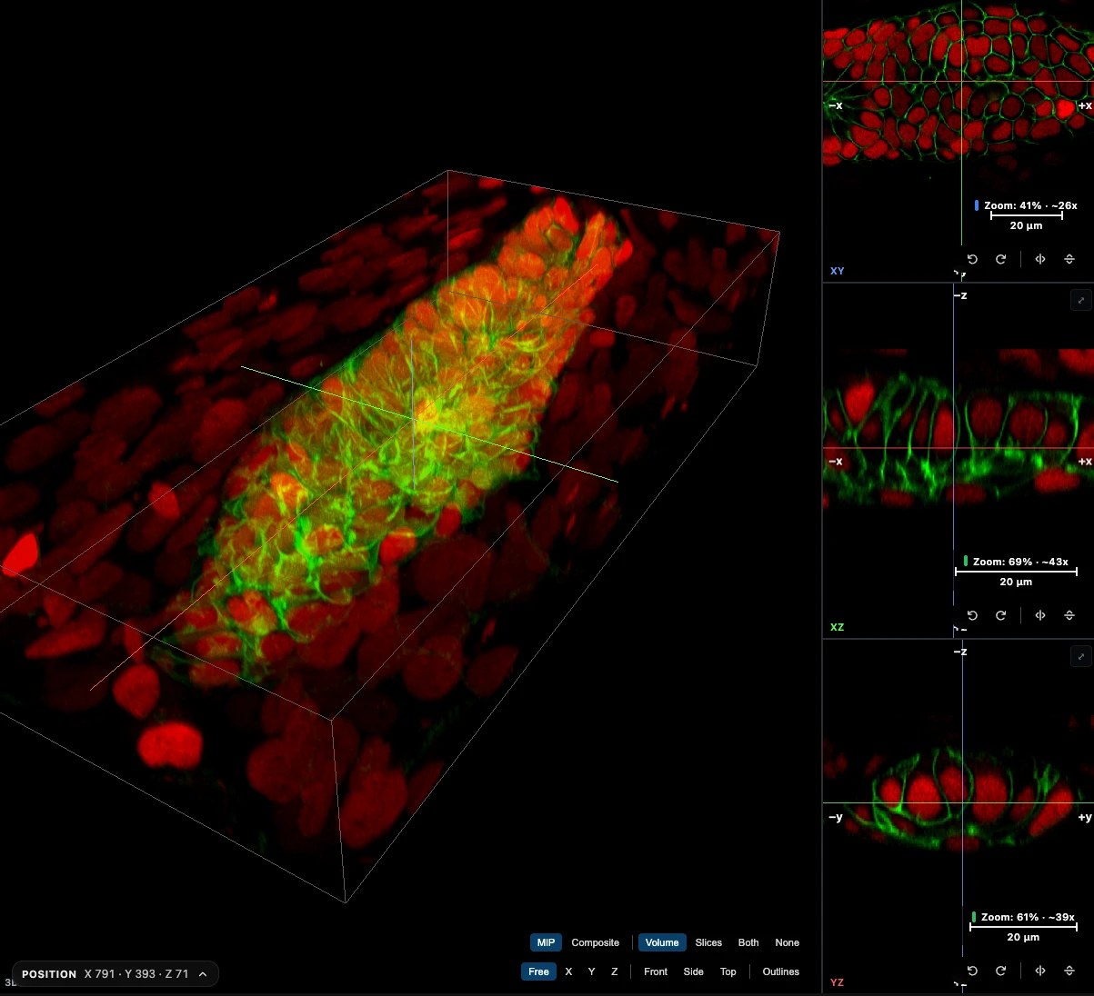
The Find Nuclei Viewer now has a 3D view. It is part of the same viewer: open an image, click 3D. Your channels, your z-plane and your annotations come with you. Free to use, nothing to install, nothing uploaded.
Try it on a real stack
The zebrafish above is the lateral line primordium from the Image Data Resource, study idr0079 [1]: 1584×788×142 voxels, membranes and nuclei.
Drag the volume to turn it. Scroll to zoom. Click MIP next to Composite to compare the two looks.
What you can do with it today
3D shows four panels: the XY plane you know, two side cuts (XZ and YZ), and the volume. The three planes share one crosshair. Click anywhere on a plane, and the other two jump to that point.
- Look at a cell from the side. Orthogonal views: put the crosshair on a nucleus, and the XZ and YZ panels show it in depth, next to XY.
- Check signal through the whole stack. Turn the volume and see where the signal fades, saturates or drifts.
- Annotate in any plane. Polygon, wand, brush, distance, points and text work on XY, XZ and YZ. Shapes show where they cross the other planes, and in the volume.
- Outline a structure over many slices. Structure mode draws one structure across slices and shows its volume in 3D.
- Go back and forth. Click 2D to return. Annotations made in either mode are the same set.
- Send a link. Copy the link in 3D, and your colleague opens the same image in 3D, with your channels and render mode.
- Keep your anatomy straight. If your image records its anatomical orientation, the axes are labelled superior and inferior, left and right, anterior and posterior, and Medical shows it the radiological way.
- Open plate fields. On an HCS plate with z-stacks, pick a well and a field, and that field opens in 3D.
MIP or Composite
Two render modes, one click apart.
MIP shows the brightest value along each line of sight. Good for bright, sparse signal: neurons, axons, puncta. It follows your contrast window, the same as 2D.
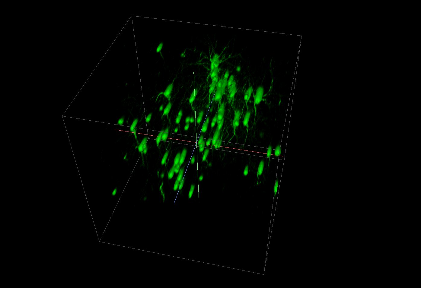
Open the marmoset neurons in 3D
Composite builds up colour and opacity front to back, so near things hide far things. Good for solid shapes. An opacity ramp per channel sets which values count as solid: raise the low end to cut haze.
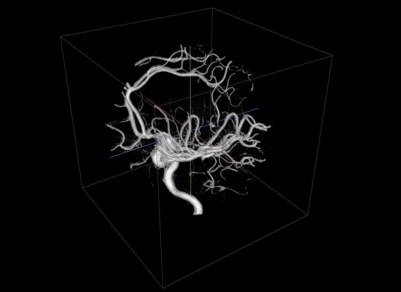
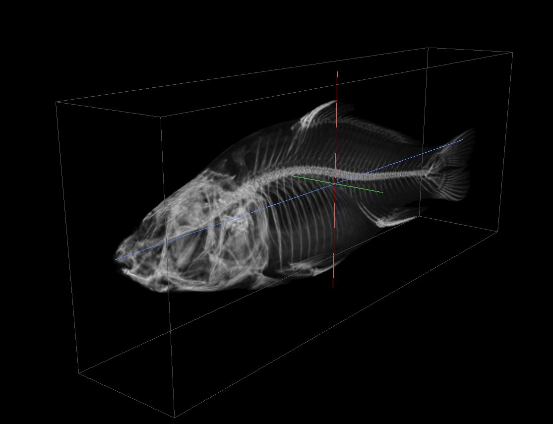
It is not only for microscopy. The gallery also has a head MRI, a skull and a carp in CT, from the OME-Zarr Open SciVis collection [2].
Bring your own stack
- Open your OME-Zarr image as you do today: from its web address, a folder on your disk, or a zipped file.
- Click 3D next to the Z slider. Or take the “Open in 3D” offer that shows when an image has z-sections.
- Look around. Click 2D to go back.
The Example Gallery has a 3D filter: seven tiles that open straight in 3D, plus the z-stacks from IDR and the BioImage Archive that you can switch to 3D yourself.
The 3D Rendering guide covers the panels, the controls, the link parameters and every limit below.
Limitations
Straight talk on limitations:
- A desktop browser with WebGPU. Recent Chrome and Edge have it. On a phone or tablet, or without WebGPU, the viewer stays in 2D.
- Four channels at once. An image can have more. Show any four.
- Big volumes come in coarser. 3D loads the whole volume at one resolution that fits in memory. A typical confocal stack shows at full detail. A whole slide or a very long stack shows coarser. The XY plane stays sharp; zoom in Planar when you need every pixel.
- Large reads take time. When 3D would read a lot of data, it tells you how much and offers to show one channel first.
- A connection. The 3D part loads the first time you click 3D. 2D works offline; 3D does not.
What comes next
Segmentation labels now show in 3D too: read Labels in 3D.
Have a z-stack that looks wrong, or does not open in 3D? Let us know at info@find-nuclei.com.
TL;DR
- Find Nuclei Viewer has a 3D view: orthogonal XY, XZ and YZ views with a shared crosshair, plus a volume rendering.
- Anatomical orientation from the image’s metadata labels the axes and drives the radiological view.
- MIP for bright, sparse signal. Composite for solid shapes, with an opacity ramp per channel.
- One click from 2D and back. Channels, z, time and annotations come with you.
- Annotate in any plane. Share links that open in 3D.
- Desktop browser with WebGPU, up to four channels at once. Opens from a URL, a local folder, an
.ozxor.zipfile, or a token-protected store. - No install, no upload. Free to use.
References
[1] Image Data Resource, study idr0079, zebrafish lateral line primordium. idr.openmicroscopy.org/study/idr0079. Paper: doi.org/10.7554/eLife.55913
[2] McCormick M. OME-Zarr Open SciVis Datasets (2025). doi.org/10.5281/zenodo.17495294. CC BY 4.0. Marmoset neurons courtesy of Frederick Federer, Moran Eye Institute, University of Utah. Aneurysm courtesy of volvis.org and Philips Research, Hamburg, Germany. Carp courtesy of Michael Scheuring, Computer Graphics Group, University of Erlangen, Germany.